Full paper available on https://arxiv.org/abs/1911.09915

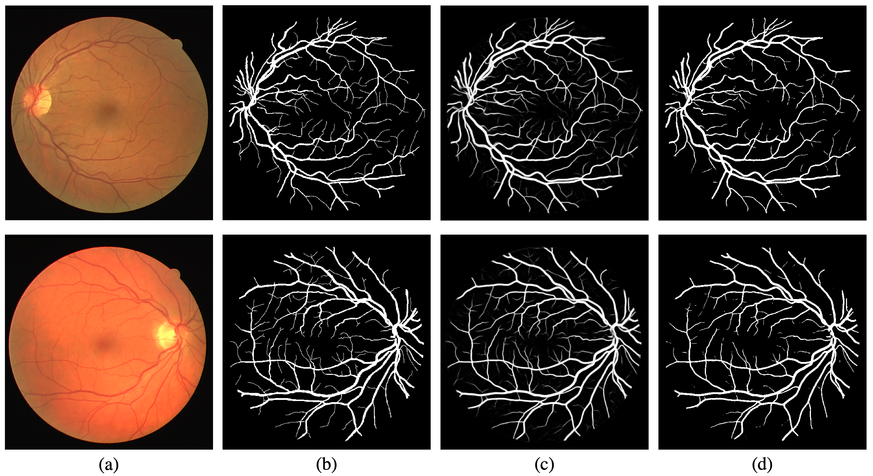
 Segmentation results for DRIVE and STARE dataset: (a) retinal image; (b) ground truth; (c) output probability map; (d) binary segmentation.
The morphological attributes of retinal vessels, such as length, width, tortuosity and branching pattern and angles, play an important role in diagnosis, screening, treatment, and evaluation of various cardiovascular and ophthalmologic diseases such as diabetes, hypertension and arteriosclerosis. The crucial step before extracting these morphological characteristics of retinal vessels from retinal fundus images is vessel segmentation. In this work, we propose a method for retinal vessel segmentation based on fully convolutional networks. Thousands of patches are extracted from each retinal image and then fed into the network, and data argumentation is applied by rotating extracted patches. Two architectures of fully convolutional networks, U-Net and LadderNet, are used for vessel segmentation. The performance of our method is evaluated on three public datasets: DRIVE, STARE, and CHASE_DB1. Experimental results of our method show superior performance compared to recent state-of-the-art methods.
- Gray-scale conversion
- Standardization
- Contrast-limited adaptive histogram equalization (CLAHE)
- Gamma adjustment
During training, 2000 patches with size 48 × 48 are randomly extracted from each image. Besides patch extraction, each extracted patch was rotated by 90◦, 180◦ and 270◦, thus the training set is quadrupled, i.e. 8000 patches are extracted from each image after data augmentation. At the end, all the patches, including original extracted and generated by rotation, are randomly shuffled and then fed into the network together with their corresponding ground truth segmentation mask patches.
During testing, images are cut into patches with size 48 × 48 and fed into the network for inference. The images are cut into patches by using a stride of N pixels (e.g. N = 5 or 10) in both height and width, so that one pixel can be predicted for multiply times. The final inference result is obtained by averaging multiple predictions.
Table 1: Segmentation results of different deep learning based methods on DRIVE. Bold values show the best score among all methods.
| Method | F1 | Sn | Sp | Acc | AUC |
|---|---|---|---|---|---|
| Melinscak et al. [1] | - | 0.7276 | 0.9785 | 0.9466 | 0.9749 |
| Li et al. [2] | - | 0.7569 | 0.9816 | 0.9527 | 0.9738 |
| Liskowski et al. [3] | - | 0.7520 | 0.9806 | 0.9515 | 0.9710 |
| Fu et al [4] | - | 0.7603 | - | 0.9523 | - |
| Oliveira et al. [5] | - | 0.8039 | 0.9804 | 0.9576 | 0.9821 |
| M2U-Net [6] | 0.8091 | - | - | 0.9630 | 0.9714 |
| R2U-Net [7] | 0.8171 | 0.7792 | 0.9813 | 0.9556 | 0.9784 |
| LadderNet [8] | 0.8202 | 0.7856 | 0.9810 | 0.9561 | 0.9793 |
| This work, U-Net | 0.8169 | 0.7728 | 0.9826 | 0.9559 | 0.9794 |
| This work, LadderNet | 0.8219 | 0.7871 | 0.9813 | 0.9566 | 0.9805 |
Table 2: Segmentation results of different deep learning based methods on STARE. Bold values show the best score among all methods.
| Method | F1 | Sn | Sp | Acc | AUC |
|---|---|---|---|---|---|
| Li et al. [2] | - | 0.7726 | 0.9844 | 0.9628 | 0.9879 |
| Liskowski et al. [3] | - | 0.8145 | 0.9866 | 0.9696 | 0.9880 |
| Fu et al [4] | - | 0.7412 | - | 0.9585 | - |
| Oliveira et al. [5] | - | 0.8315 | 0.9858 | 0.9694 | 0.9905 |
| R2U-Net [7] | 0.8475 | 0.8298 | 0.9862 | 0.9712 | 0.9914 |
| This work, U-Net | 0.8219 | 0.7739 | 0.9867 | 0.9638 | 0.9846 |
| This work, LadderNet | 0.7694 | 0.7513 | 0.9764 | 0.9529 | 0.9660 |
- numpy >= 1.11.1
- PIL >=1.1.7
- opencv >=2.4.10
- h5py >=2.6.0
- ConfigParser >=3.5.0b2
- scikit-learn >= 0.17.1
Training from scratch:
python run_training.py <configuration_drive.txt|configuration_stare.txt|configuration_chase.txt>
Keep training based on pretrained models:
python run_keep_training.py <configuration_drive.txt|configuration_stare.txt|configuration_chase.txt>
python run_testing.py <configuration_drive.txt|configuration_stare.txt|configuration_chase.txt>
[1] Melinscak, M., Prentasi, P., & Lonari, S. (2015, January). Retinal vessel segmentation using deep neural networks. In VISAPP 2015 (10th International Conference on Computer Vision Theory and Applications).
[2] Li, Q., Feng, B., Xie, L., Liang, P., Zhang, H., & Wang, T. (2016). A Cross-Modality Learning Approach for Vessel Segmentation in Retinal Images. IEEE Trans. Med. Imaging, 35(1), 109-118.
[3] Liskowski, P., & Krawiec, K. (2016). Segmenting retinal blood vessels with deep neural networks. IEEE transactions on medical imaging, 35(11), 2369-2380.
[4] Fu, H., Xu, Y., Lin, S., Wong, D. W. K., & Liu, J. (2016, October). Deepvessel: Retinal vessel segmentation via deep learning and conditional random field. In International Conference on Medical Image Computing and Computer-Assisted Intervention (pp. 132- 139). Springer, Cham.
[5] Oliveira, A. F. M., Pereira, S. R. M., & Silva, C. A. B. (2018). Retinal Vessel Segmenta- tion based on Fully Convolutional Neural Networks. Expert Systems with Applications.
[6] Laibacher, T., Weyde, T., & Jalali, S. (2018). M2U-Net: Effective and Efficient Retinal Vessel Segmentation for Resource-Constrained Environments. arXiv preprint arXiv:1811.07738.
[7] Alom, M. Z., Hasan, M., Yakopcic, C., Taha, T. M., & Asari, V. K. (2018). Recurrent Residual Convolutional Neural Network based on U-Net (R2U-Net) for Medical Image Segmentation. arXiv preprint arXiv:1802.06955.
[8] Zhuang, J. (2018). LadderNet: Multi-path networks based on U-Net for medical image segmentation. arXiv preprint arXiv:1810.07810.


