start=>start: 开始
operation1=>operation: image split 成1024 * 1024
operation2=>operation: image transfer
operation3=>operation: image deconvolution 得到He
operation4=>operation: open morphological operation
operation5=>operation: close morphological operations
operation6=>operation: Otsu's thresholding
operation7=>operation: open morphological operations
operation8=>operation: remove small area and small holes
operation9=>operation: remove artifact
operation10=>operation: run adaptive multi-scale LOG filter
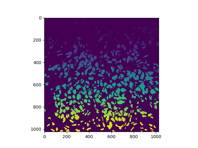
operation11=>operation: detect and segment nuclei using local maximum clustering
operation12=>operation: filter out small objects
start->operation1->operation2->operation3->operation4->operation5->operation6->operation7->operation8->operation9->operation10->operation11->operation12
-
image transfer一定要选取对比度鲜明的图像作为模板
-
开闭操作一定选择椭圆核,这样可以得到更平滑的图像,而且得到的结果更倾向于椭圆,符合我们的要求
-
移除小孔和识别伪影这里都是设置了面积的阈值,目前没有更好的方法
-
首先需要确定三个超参数:min_radius、max_radius、local_max_search_radius。我现在生成了三个超参数不同组合的图片,共有近千张,需要选出最优超参数。我建议找刘主任标注出来一张图像,然后进行比对。
-
粗分割时可能会带有一些细胞质进来,导致分割结构不规则,在后面的细分割得到一些奇怪的结构
-
对于伪影的处理,使用控制面积阈值的方法其实并不靠谱,希望可以有更优的方法
-
得到细分割的结果之后,对每个细胞进行统计半径,面积,长宽比等特征,进行分类
-
算法加速:建议使用Cython重构一下,我之前写过,出了点问题,就删掉了
-
可以了解一下病理学知识,还有生物统计学
-
可以写一个带有界面的勾画软件,把每次得到的结果发给刘主任,让他们使用软件进行修改,多次循环后得到的结果就可以用于深度学习训练了