Topics
Theory
- Light
- Frequency, wavelength, and energy
- Polarity
- Wave mechanics
- The photoelectric effect
- Absorption and emission -- Stokes shift
- Lenses
- Snell etc
- Thin-lens
- Manufacturing
- Photon sieves
- Sensors
- CMOS
- APD
- PMT
- Consequences
- Depth of field
- Airy disks
- Resolution
- Chromatic abberation
- Photo-bleaching
Instrument Application
- Basics
- Microscope anatomy
- Objectives
- Parameters
- Metadata
- Slides and Coverslips
- Polylysine
- https://www.genomv.wis3.edu/pub/reprints/GEC_Slide_Coating.pdf
- CC2
- Soda lime
- Borosilicate 1.0 / 1.5 (high-borate transmits UV)
- Permanox plastic
- RS
- Silicone
- PEN membrane glass
- Cell-culture
- Nuclon Delta
- White-light
- Bright-field
- Dark-field
- Phase-contrast
- Differential-interference contrast
- Fluorescent
- Dyes
- Titration
- Detection
- Quantification
- Advanced Techniques
- Confocal
- Light-sheet
- Multi-photon
- STORM
Sample Application
- Immunohistochemical
- Dyes
- Labels
- Chemistry
- Immunofluorescent
- Dyes
- Labels
- Panel design
- Preparation
- Cryogenic
- Paraffin-embedded
- Storage
- Efficiency
- Automated tools
Analysis
- ImageJ
- Resources
- Recording and building macros
- Standardizing a pipeline
- Quantitation
- Masking
- Strategies for labeling objects
- Presentation
- Pipeline to plots
- Displaying color clearly
- Avoiding bias
Using a maximum likelihood estimation (MLE) reconstruction algorithm for single point-spread function fitting.
https://andor.oxinst.com/learning/view/article/comparing-scmos
https://www.photometrics.com/learn/super-resolution-microscopy
Super-Resolution Microscopy
Super-resolution microscopy techniques are so-called because of their ability to resolve structures beyond the diffraction limit of light. Conventional light microscopy techniques are unable to bypass this limit, which prevents structures finer than roughly half the wavelength of the emission light (typically no less than ~200 nm) from being resolved. In contrast, super-resolution techniques have been shown to achieve single nanometer resolution.
https://www.photometrics.com/learn/spinning-disk-confocal-microscopy/what-is-super-resolution-microscopy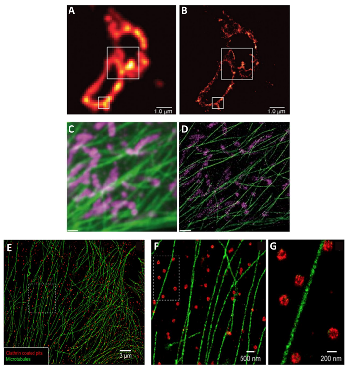**Figure 3: Images from different super-resolution** localization techniques, compared to standard microscopy. A) Standard microscopy and B) PALM images of transmembrane proteins (image from Betzig et al. 2006). C) Standard microscopy and D) DNA-PAINT images of mitochondria (magenta) and microtubules (green) from within a cell (image from Jungmann et al. 2014). E/F/G) Two-color STORM images of microtubules (green) and clathrin-coated pits from within a mammalian cell at increasing magnifications, the area indicated by dashed box on the previous image (image from Bates et al. 2007).
