End-to-end PSF extraction for 3D microscopy
This is a project on non-parametric estimation of 3-dimensional shape of Point Spread Function (PSF) of an optical system with the help of super-resolution simgle image generative adversarial network (SRGAN).
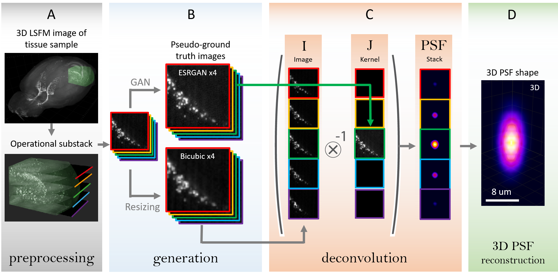
This project is aimed at resolution enhancement of light-sheet fluorescent microscopy images of a large tissue samples (whole organs, bodies). The pseudo-ground truth images are generated by GAN. These images are used as kernels in deconvolution for the point spread function (PSF) estimation. The further 3D deconvolution with these PSFs of a bicubically upsampled image allows to increase the image resolution with no fake features generation.
In the current project the Enhanced SRGAN is used from the original mmsr github repo. The original article by Wang et al., discribing the architecture, training process and weight interpolation process is available on arxiv.
The used GAN architecture in application to 3D stack of microscopy images:
ESRGAN architecture
- PyTorch (An open source deep learning platform)
If for some reason you choose not to use Anaconda, you must install the following frameworks and packages on your system:
- Python 3.7
- Pytorch >= 1.0 (CUDA version >= 7.5 if installing with CUDA)
- tifffile - package to work easily read and save 16-bit .tiff files from microscopes
- opencv >= 4.1.0
- imutils - simple opencv add-on
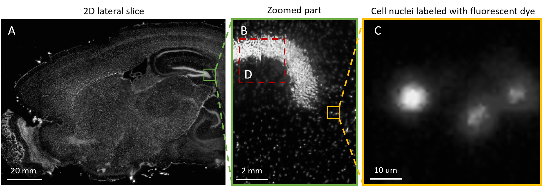
The used data represents the 3D images imaged with the light-sheet fluorescent microscopy in collaboration with Brain Stem Cell Lab, MIPT & CSHL. The data is huge enough, so it is located at Computational Imaging Group local servers. There are 2 Datasets:
Available at cig7-server/roman_kiryanov/data-Z1/
Represents a part of mouse brain with cell nuclei labeled by propidium iodide (PI). This dataset is used for GAN fine-tuning.
Dataset 1
Available at cig7-server/roman_kiryanov/data-LightSheet/
Represents a whole mouse brain with cell nuclei labeled by 5-Ethynyl-2'-deoxyuridine (EdU). This dataset is used for GAN validation and PSF estimation.
Dataset 2
Both datasets are unsigned 16 bit grayscale image .tiff stack. In microscopy_data_classes.py the useful classes for data manipulation are present. They allow to load, visualize and crop the 3D volumes.
There are 3 main stages in this work. The first one is ESRGAN fine tuning, the second is a deconvolution with pseudo-generated images for PSF estimation, and the final is a 3D deconvolution of images with extracted PSFs.
The general idea of the proposed method consist of several parts:
PSF estimation stages
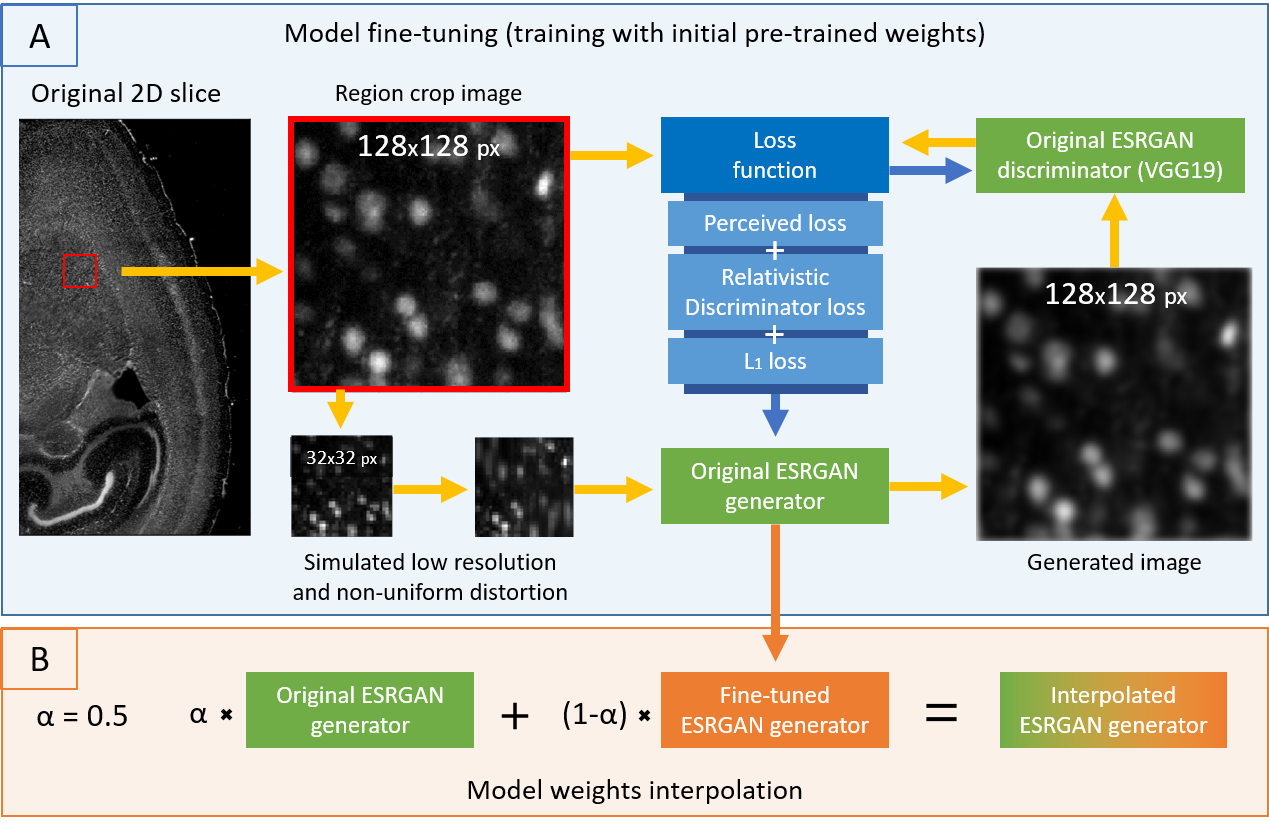
The code for data generation for GAN training is all combined in a one notebook eSRGAN_data_generation_for_training.ipynb. There the ste-by-step process of generation training data from lateral slices of original Dataset 1 is present. The code for ESRGAN fine0tuning is available in notebook eSRGAN_manipulations_Jan2020.ipynb. For the simplicity, the most necessary part for ESRGAN frine-tuning of mmsr github repo is copied in the folder ./mmsr_code/.
The pipeline for GAN fine-tuning:
ESRGAN optimization pipeline
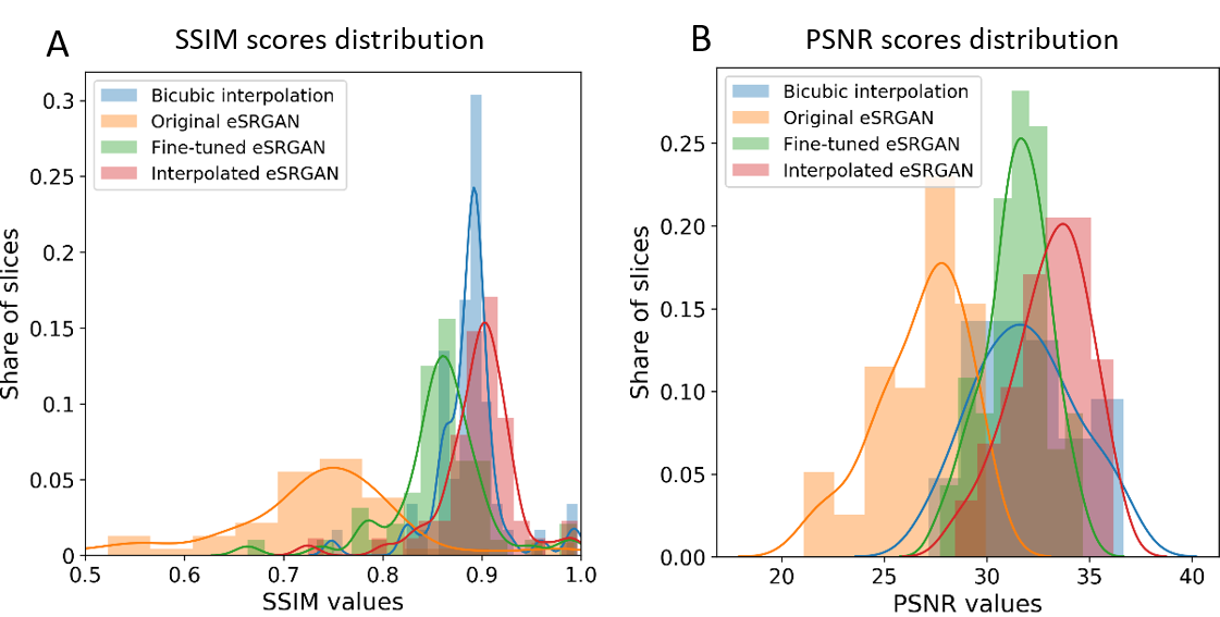
The results on test subset of Dataset 1 were measured with PSNR and SSIM metrics. The file distributions_psnr_ssim_dataset_1.pkl is a simple Python pickle object that contains these distributions:
Generation comparison on test subset of Dataset 1
The proposed method for PSF estimation is available in notebook eSRGAN_results_evaluation_and_hypothesis_tests.ipynb.
the general approach is described im Thesis, while shortly, the generated by GAN images are used as kernels in nin-blimd deconvolution (in the present work Unsupervised Wiener filter from scipy was used).
The resulting data of some experiments is available in the folder ./data/ of the current repo.
- In subfolder
psf_extraction_synthetic_data_05052020the encoded and extracted PSFs from the experiment on synthetic data are stored in a form of .tiff 3D stacks. - The code for all experiments is present in
Synthetic_data_manipulation.ipynb
- In subfolder
psf_extraction_real_data_operational_substack_LS_03052020the extracted by several methods PSFs are stored in a form of .tiff 3D stacks. - In notebook
PSF_analysis_29052020.ipynbthe analysis of extracted PSFs is given with collected metrics on deconvolved images.
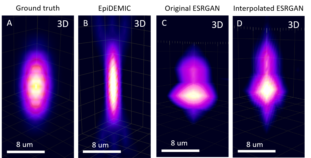
Extracted PSFs:
Extracted PSFs
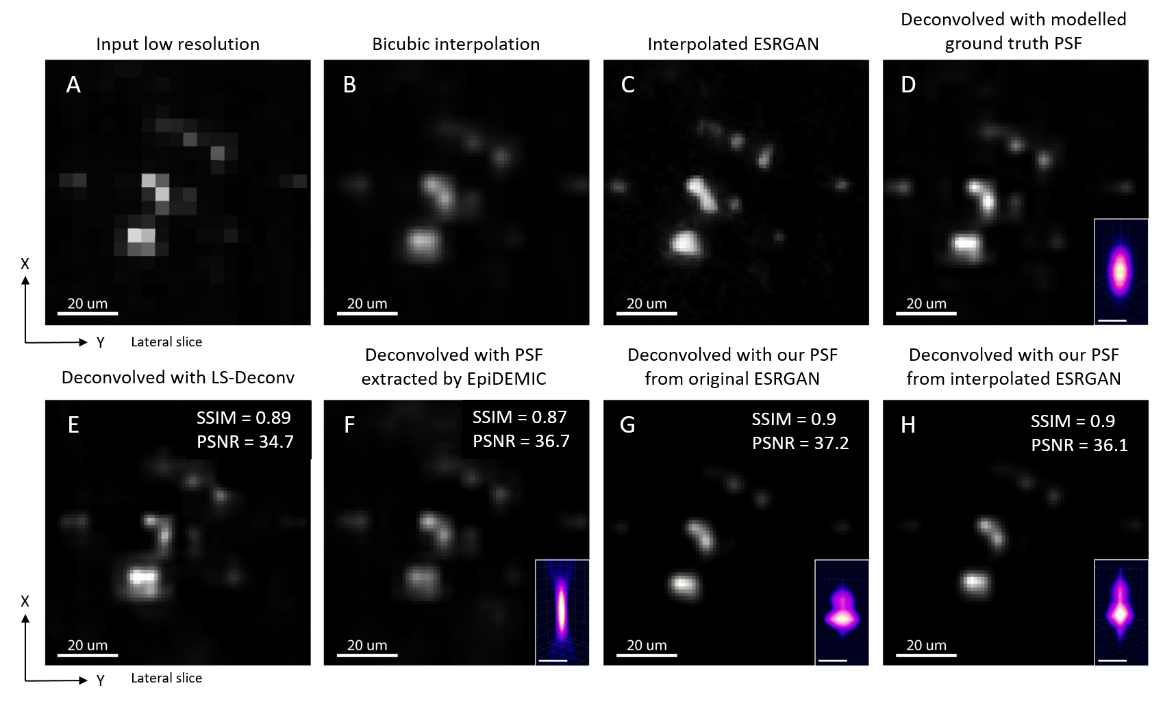
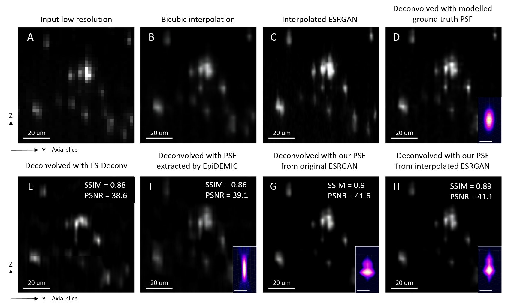
This stage was done outside of Python, in ImageJ plugin DeconvolutionLab2. The Richardson-Lucy deconvolution with 10 iterations and
Results on lateral slice:
Results on lateral slice
Results on axial slice:
Results on axial slice
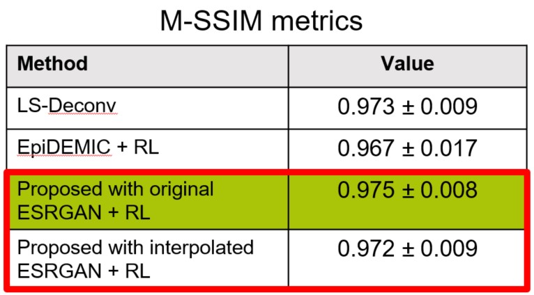
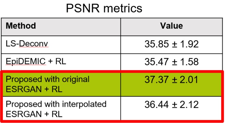
Numerical comparison of the results The used GAN architecture in application to 3D stack of microscopy images:
roman.kiryanov@skoltech.ru, Roman Kiryanov


