Image Segmentation of Brain Tumors using Convolutional Neural Networks
In this project we implement two different networks in order to create segmentation masks for identifying brain tumors.
The complete dataset can be accessed here
Overview
Dataset Used
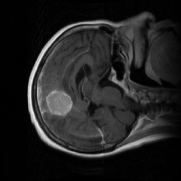
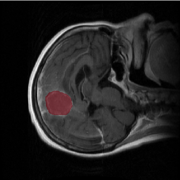
This brain tumor dataset contains 3064 T1-weighted contrast-inhanced images from 233 patients with three kinds of brain tumor: meningioma (708 slices), glioma (1426 slices), and pituitary tumor (930 slices). Each image has the dimension (512 x 512 x 1). The full dataset is available here
| Original Image | Original Image With Mask |
|---|---|
 |
 |
Model Architecture
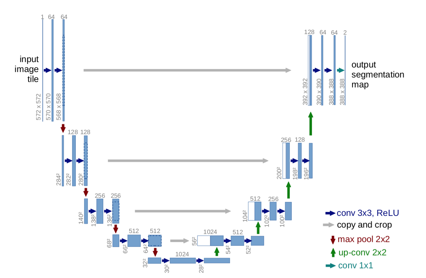
The model architecture for U-Net
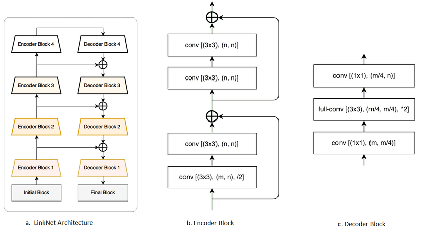
The model architecture for LinkNet
Training Process
The networks were trained on a system with an AMD Ryzen 5 2600 processor overclocked to 4.0GHz, 16GB of RAM and an NVIDIA GeForce RTX 2070 GPU.
U-Net
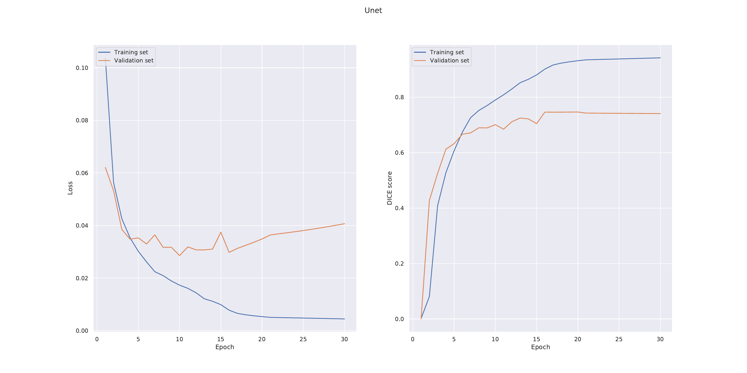
We experimented with the number of initial filters and settled for 16. U-net was found to perform well when using a batch size of 8. The initial learning rate was set to 10e−4 .
LinkNet
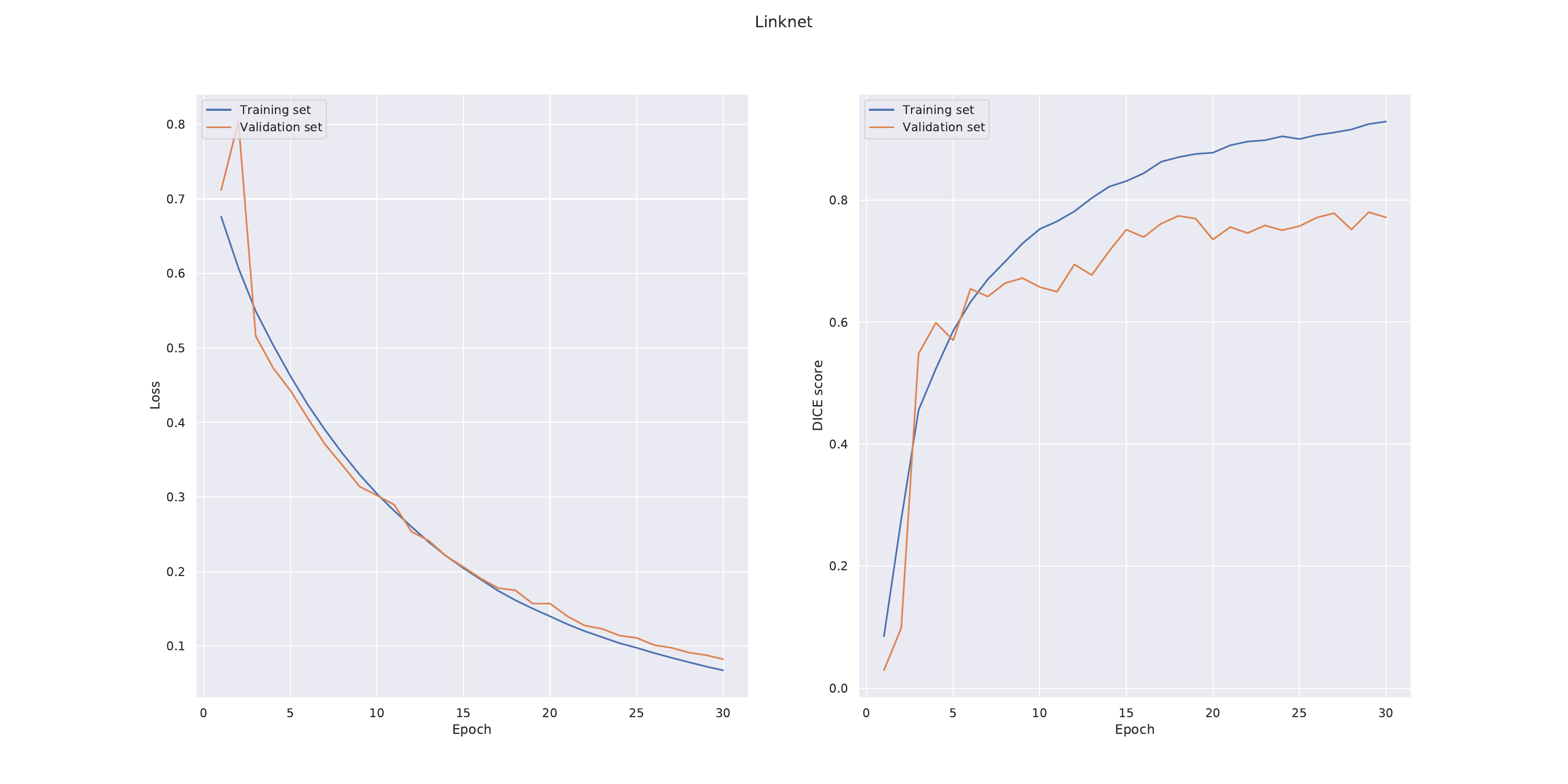
We experimented with the number of start filters and found that 64 seemed to give the best results. We noticed that LinkNet seemed to be sensitive to parameter settings. LinkNet was trained using an initial learning rate of 5 · 10e−4 and a batch size of 10. Batch normalisation was used after each convolutional layer. We also tried adding L2-regularisation but that did not seem to significantly improve training.
Results
The final DICE scores achieved by the respective networks on the test set were
| Method | DICE Score |
|---|---|
| U-Net | 0.703 |
| LinkNet | 0.715 |
Some samples from the predictions are shown below
| U-Net | Linknet |
|---|---|
 |
 |
 |
 |
 |
 |
 |
 |
 |
 |
